- 王蕙丽文章图片
- 来源:于明明教授个人网站 2023-02-22

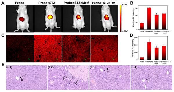
(A) In vivo imaging of DPX in mice after different stimulations: mice treated with the probe only, mice pretreated with STZ, mice pretreated with STZ and further treated with Metf, and mice pretreated with STZ and further treated with RVT. (B) Relative fluorescence intensity of mice corresponding to A. (C) Fluorescence images of DPX in various liver tissues corresponding to A. (D) Relative fluorescence intensity of tissues corresponding to A. (E) H&E staining images of liver tissues from mice treated with the probe only, probe + STZ, probe + STZ + Metf, and probe + STZ + RVT, respectively.
- [来源:中国聚合物网]
- 了解更多请进入: 于明明教授个人网站